Non-surgical rhinoplasty is a popular cosmetic procedure that involves the use of injectable fillers to improve the shape of the nose without surgery. There are many different materials available on the market such as hyaluronic acid, collagen stimulators, etc. Silicone has also been used in non-surgical rhinoplasty, however, it is not Food Drug Administration (FDA)-approved due to the nature of the material and the higher risk of complications, such as infection, secondary inflammation, deformities, granulomas, and scar tissue formation.
Surgical removal has always been the only way to remove silicone-based filler injection. However, it has a long recovery time and can cause extensive scar tissue and deformation. Foreign body granuloma can occur with silicone injection and often intralesional steroid injection is used to deal with this granuloma.
Case report
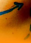
A 38-year-old female attended the clinic with hypopigmentation at nasion for the last five years. According to the patient, she had a silicone-based material injected into her nose a decade ago. Due to the nose looking unnatural following this procedure, she went to a doctor and received an intralesional triamcinolone injection as a method to deal with the silicone material after failing with hyaluronidase injection. The steroid injection did help with reducing the swelling, however, hypopigmentation with a ring of hyperpigmentation formed not long after. Since then, the patient tried multiple sessions of platelet rich plasma injection but to no avail. She requested lightening the ring of hyperpigmentation and possibly restoring some pigment to the hypopigmented skin.
The treatment plan was to lighten the hyperpigmentation area with the Nd:YAG picosecond laser and attempt to restore pigment to the hypopigmented area with polynucleotides. The patient’s expectations were managed prior to the treatment being undertaken.

EMLA cream was applied to the affected area for 40 minutes prior to the treatment. The first step was to cover the hypopigmentation area with white colour plaster. The pigmented area was treated with Fotona StarWalker PQX, black handpiece with a 4mm spot size, fluence 3J/cm2. Multiple passes were completed with no pain reported. We followed this by using the black F5 handpiece, fluence 2J/cm2, and the endpoint of mild erythema was achieved in the same area.
The last step was to inject polynucleotides intradermally into the hypopigmented area using the nappage injection technique. There were a few small bumps after injection, which subsided within one to two days. Moisturiser and sunscreen were applied to the area after treatment. The patient was advised to avoid sun exposure.
The patient returned for follow-up after one month and was satisfied with the result after only one session. No complications were observed. With a subsequent follow-up visit at four months post-treatment, the results have been sustained with no regression noted. In this patient, the combination of Nd:YAG laser and polynucleotides treatment proved an efficient way to deal with iatrogenic dyspigmentation caused by intralesional steroid injection.
Discussion
Subcutaneous fat atrophy, telangiectasia and hypopigmentation are well-known side-effects of steroid injection. In this case, steroid injection did help with the reduction of swelling, however, the procedure left a rather unpleasant appearance for the patient causing her to experience stress and a lack of self-confidence.
The mechanism of the hypopigmentation is not yet known. However, there is a ring of hyperpigmentation around the depigmented area, this could be the result of inflammation following steroid injection.
Polynucleotides consist of DNA derived from the sperm cells of salmon and trout. In this case, polynucleotides restored the damaged micro-environment, hence rebuilding the new melanocyte and melanin at the target area [1]. To conclude, the combination of laser and polynucleotide injection is an effective tool to treat iatrogenic dyspigmentation caused by steroid injection.
References
1. Khan A, Wang G, Zhou F, et al. Polydeoxyribonucleotide: a promising skin anti-aging agent. Chin Jour of Plas and Recon Surg 2022;4(4):187–93.
2. Lee JM, Kim YJ. Foreign body granulomas after the use of dermal fillers: pathophysiology, clinical appearance, histologic features, and treatment. Arch Plast Surg 2015;42(2):232–9.
Declaration of competing interests: None declared.
COMMENTS ARE WELCOME


