This article has been verified for CPD. Click the button below to answer a few
short questions and download a form to be included in your CPD folder.
Post-inflammatory hyperpigmentation (PIH) secondary to any inflammatory cause or cutaneous injury is a common complaint [1-2]. There are significant quality-of-life and psychological implications [3] particularly for patients with non-white skin, due to its chronic and unpredictable course [1]. It is characterised by an abnormal and increased distribution of melanin which often lingers for months after the initial insult to the skin. [1].
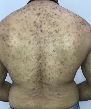
In this case study, I share the management of active back acne and severe PIH in a 27-year-old Indian man. The acne had persisted for over 10+ years. The patient had been seen previously by his GP and a dermatologist. There were compliance issues with topical medication (including salicylic acid) and two courses of Roaccutane were unfinished due to intolerable dryness. He was not keen to repeat this experience. His only skincare was washing with an emollient antiseptic soap substitute. Of note he had used a tight and heavy backpack daily on a long commute over the last 10 years. On examination (Figure 1), the patient had oily, textured skin with a thickened stratum corneum and active acne lesions which he frequently picked. He had excess hair growth which he removed regularly with waxing. Examination with a Woods lamp demonstrated a mixed picture of epidermal and dermal hyperpigmentation.

Figure 1.
Acne vulgaris is a chronic condition of the pilosebaceous unit characterised by an overproduction of oil, inflammation and proliferation of C. acnes bacteria [4]. For optimum patient outcomes and to minimise PIH, treatment regimens including lifestyle changes must control all of these processes [4].
Melanogenesis occurs in melanosomes as a protective mechanism following inflammation in skin of colour [2]. Tyrosinase is the rate-limiting enzyme in the cascade of events in which tyrosine is oxidised to dopa and dopa is oxidised to melanin [5-7]. Melanogenesis, melanin transfer and melanin distribution all impact the presence of hyperpigmentation in the skin and treatment modalities should interfere at different steps to improve patient outcome.
In this case, exfoliation was key to releasing underlying congestion and preventing pseudofolliculitis barbae. The homecare regime included three steps twice daily: 1.18% glycolic acid cleanser with lactobionic acid to improve toleration; 2. A gentle hypochlorous antimicrobial solution (to be used as a spray for its ease of application on difficult to reach areas of the back) to combat the purge following intense exfoliation; and 3. 10% glycolic acid lotion with citric acid for its potent antioxidant and skin brightening properties [8].
Glycolic acid, an alpha hydroxy acid has the ability to modulate both epidermal and dermal changes at higher concentrations [8]. It facilitates renewal of the stratum corneum by reducing corneocyte cohesion and modulates desmosomal attachments between corneocytes improving skin texture and flexibility of the skin barrier [9].
Glycolic acid prevents pore occlusion and also has bactericidal properties [4]. It potently inhibits C. acnes by disrupting cell membranes in the pH range of 3-4.5 [4], which is compatible with homecare preparations. It also reduces melanin clumping and the appearance of hyperpigmentation [1,9,10] with well documented safety for use in all Fitzpatrick skin types [11]. Superficial exfoliation with alpha hydroxy acids in homecare and chemical peels improves the delivery of pigment modulating agents [10]. It is also preferred for darker skin types compared to medium depth peels which impact the papillary dermis and deep peels which impact the reticular dermis as these can cause more irritation, a precursor to further PIH [10].
An extended acclimatisation period was essential given the darker skin type to minimise dermatitis. After one month the acne lesions and underlying congestion had reduced, skin texture was smoother and PIH was beginning to improve with an overall brighter tone. At this point the patient underwent a layered peel with a 30% mandelic and citric acid followed by 20% glycolic acid. A superficial retinol peel solution was then applied for 12 hours. The peel was controlled to ensure the patient did not experience any irritation. His aftercare included the antimicrobial solution and a lactobionic acid moisturiser with humectant and antioxidant benefits to condition and heal the skin barrier after which he returned to his pre-peel regimen.
The patient’s second peel was planned to take place six weeks later, however 2020 brought new challenges to clinical practice with COVID-19 and lockdown restrictions causing disruption. At this point, 10 weeks into treatment the acne lesions had significantly reduced. The patient was keen to continue treatment, therefore, cysteamine (5%) was introduced daily. At night the patient continued his existing regime. Photography was submitted monthly to review progress.
Cysteamine is a naturally occurring aminothiol physiologically produced in human cells found to induce 80% melanin synthesis reduction in vitro [12]. It has a multi-fold mechanism of action on the melanogenesis cascade through tyrosine inhibition, peroxidase inhibition, iron and copper quenching, dopaquinone quenching. It increases intracellular glutathione and activates the phaeomelanin pathway, it inhibits melanosome transfer and also lightens epidermal melanin in the stratum corneum via a potent antioxidant effect [13-15]. Increasing glutathione is an important mechanism of action as depletion of glutathione has been shown to increase tyrosinase activity [16] and glutathione is also thought to have a dose dependant inhibition on melanin synthesis [5]. The efficacy of cysteamine has been extensively tested in darker skin tones with a study by Mansuri et al. receiving the Heinz Maurer award in 2017 for research in the ethnic population [14]. It has also been proven to have better efficacy and tolerability than triple therapy with Kilgman Formula [15].

Figure 2.
After one month a significant improvement in PIH was noted (Figure 2) and cysteamine was continued for another three months. As the acne had improved, the patient admitted to reduced compliance with his antimicrobial solution, and that he was picking the skin more due to stress during the final month. Despite this, there was further improvement in his PIH. A twice-weekly maintenance dose of cysteamine was then continued alongside his existing regime. Seven months later than originally planned, the patient continued with two further layered peels as per the original treatment plan.

Figure 3.
He was extremely happy with his results (Figure 3), especially that he was able to continue treatment with remote support. He reports that the skin feels much healthier and smoother and that his confidence has grown. He will commence laser hair removal and continues with cysteamine maintenance and his antimicrobial wash. We will initiate next steps including micro-needling thereafter.
References
1. Shenoy A, Madan R. Post-inflammatory hyperpigmentation: a review of treatment strategies. J Drugs Dermatol 2020;19(8):763-8.
2. Halder RM, Grimes PE, McLaurin CI, et al. Incidence of common dermatoses in a predominantly black dermatologic practice. Cutis 1983;32:388-90.
3. Lacz NL, Vafaie J, Kihiczak NI, et al. Post inflammatory hyperpigmentation: a common but troubling condition. Int J Dermatol 2004;43:362-5.
4. Valle-Gonzalez ER, Jackman JA, Yoon BK, et al. pH-Dependent antibacterial activity of Glycolic Acid. Implications for anti-acne formulations. Sci Rep 2020;10:7491.
5. Kligman AM, Grove GL, Hirose R, Leyden JJ. Topical tretinoin for photo-aged skin. J Am Acad Dermatol 1986;15:836-859.
6. Chaudhuri R. Hexylresorcinol: Providing Skin Benefits by Modulating Multiple Molecular Targets. In: Sivamani RK, Jagdeo JR, Elsner P,Maibach HI (Eds). Cosmeceuticals and Active Cosmetics Third Edition CRC Press; 2016;71-82.
7. Makino ETT, Mehta RCC, Banga A, et al. Evaluation of a hydroquinone free skin brightening product using invitro inhibition of melanogenesis and clinical reduction of ultraviolet-induced hyperpigmentation. J Drugs Dermatol 2013;12(3):16-20.
8. Rendon MI, Berson DS, Cohen JL, et al. Evidence and considerations in the application of chemical peels in skin disorders and aesthetic resurfacing. J Clin Aesthetic 2010;3:32-43.
9. Van Scott EJ, Yu RJ. Hyperkeritinization, corneocyte cohesion and alpha hydroxy acids. J Am Acad Dermatol 1984;11(5):867-79.
10. Chaowattanapanit S, Silpa-Archa N, Kohli I, et al. Postinflammatory hyperpigmentation: A comprehensive overview. Treatment options and prevention. J Am Acad Dermatol 2017;77:607-21.
11. Sharad J. Glycolic acid peel therapy –current review. Clin Cosmet Investig Dermatol 2013;6:281-8.
12. Qiu L, Zhang M, Sturm RA, et al. Inhibition of melanin synthesis by cystamine in human melanoma cells. J Invest Dermatol 2000;114(1):21-7.
13. Austin E, Nguyen JK, Jagdeo J. Topical treatments for Melasma: A systematic Review of Randomized Controlled Trials. J Drugs Dermatol 2019;18(11):1156.
14. Mansouri P, Farshi S, Hashemi Z, Kasraee B. Evaluation of the efficacy of cysteamine 5% cream in the treatment of epidermal melasma: a randomiased double-blind placebo controlled trial. BJD 2015;173(1):209–17.
15. Kasraee B, Mansouri P, Farshi S. Significant therapeutic response to cysteamine cream in a elasma patient resistant to Kligman’s formula. J Cosmet Dermatol 2019;18(1):293-5.
16. del Marmol V, Solano F, Sels A, et al. Glutathione depletion increases tyrosinase activity in human cells. J Invest Dermatol 1993;101:871-4.
Declaration of competing interests: The author is a Key Opinion Leader and Trainer for Aesthetic Source.
COMMENTS ARE WELCOME


